By A Mystery Man Writer

Download scientific diagram | Medial view of left knee region highlighting various fascial components surrounding the semitendinosus muscle. From the superficial to the deep aspect: the fascia lata, the paratenon and the epimysium from publication: Anatomical study of paratenons and fascia lata connections in the posteromedial knee region | Introduction In the last decade, fascia research increased significantly in various aspects such as anatomical and biomechanical features related to epimuscular force transmission. Methods The present anatomic study focuses on macroscopic observations of the potential | Fascia Lata, Hamstring muscles and Fascia | ResearchGate, the professional network for scientists.
Right knee lateral and meniscus depicts the Medial Femoral Condyle, Lateral Femoral Condyle, superior view of meniscus and tibial plateau.

Normal anatomy of the right knee

Anatomical study of paratenons and fascia lata connections in the posteromedial knee region

Marcel ROOZE, Université Libre de Bruxelles, Brussels, ULB, Laboratory of Anatomy, Biomechanics and Organogenesis
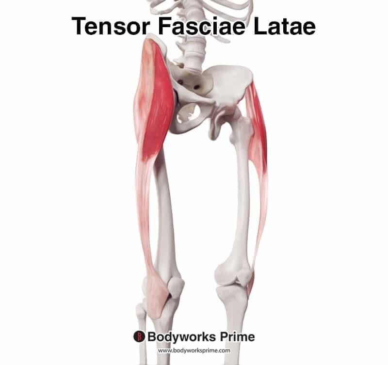
Tensor Fasciae Latae (TFL) Muscle Anatomy - Bodyworks Prime

1. Rana esculenta. (A) Pelvis (dorsal view); fascia removed on

Benoit BEYER, Assoc. Prof., PT, MSc, PhD, Université Libre de Bruxelles, Brussels, ULB, Faculty of Motricity Sciences (FMS)
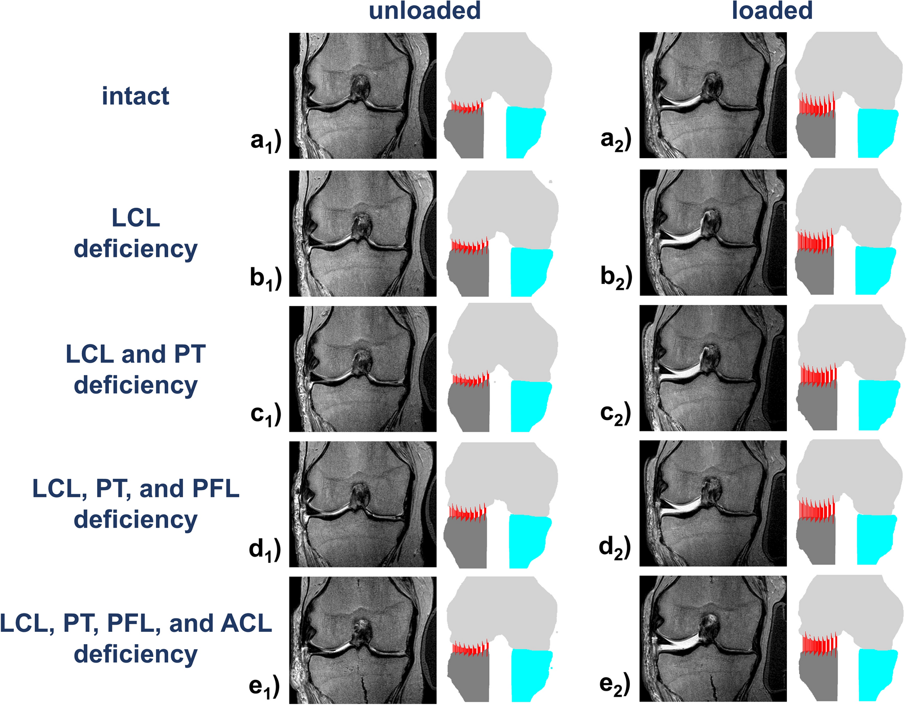
Varus stress MRI in the refined assessment of the posterolateral
Applied Sciences, Free Full-Text
IJERPH, Free Full-Text
Knee - Physiopedia

Anatomy of the posterior aspect of a right knee with the medial

Fascial Manipulation Practical Part: Luigi Stecco, Carla Stecco
/images/vimeo_thumbnails/258787002/P5TcgBAWlKmMMkIh5VQ_overlay.jpg)
Middle ear: Anatomy, relating structures and supply

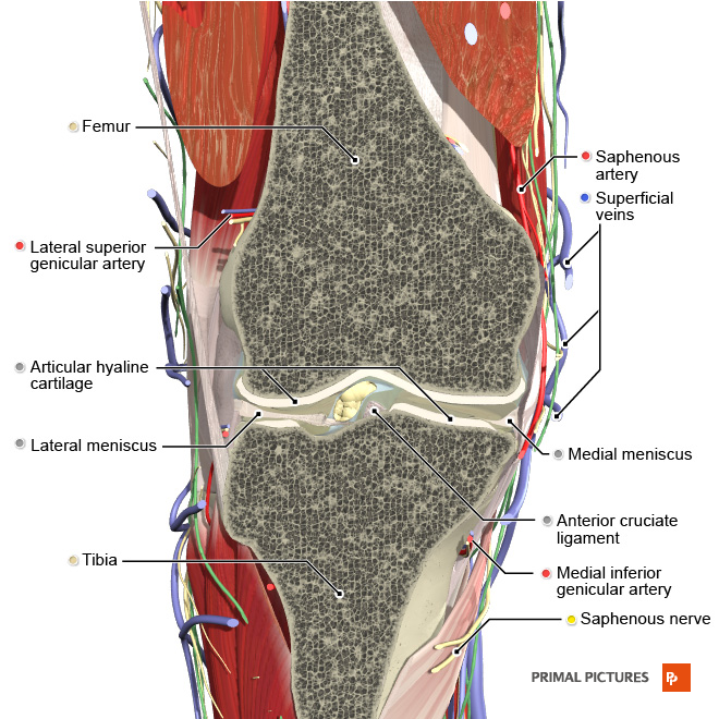
Knee Joint Cross Section - Medical Art Library

AP and lateral radiographs of the left knee demonstrate lucency