By A Mystery Man Writer

Given the twelve thoracic vertebrae are largely similar, most are considered typical thoracic vertebrae with the exceptions T1 and T9 to T12. For a basic anatomic description of the structure of ty

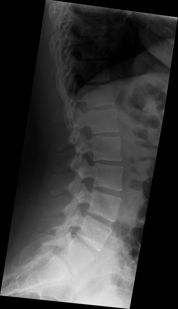
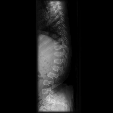
Thoracolumbar spine x-rays - Don't Forget the Bubbles

Spondylosis, Radiology Reference Article
A and B) Posteroanterior and lateral X-ray image of the thoracic

Thoracolumbar spine x-rays - Don't Forget the Bubbles

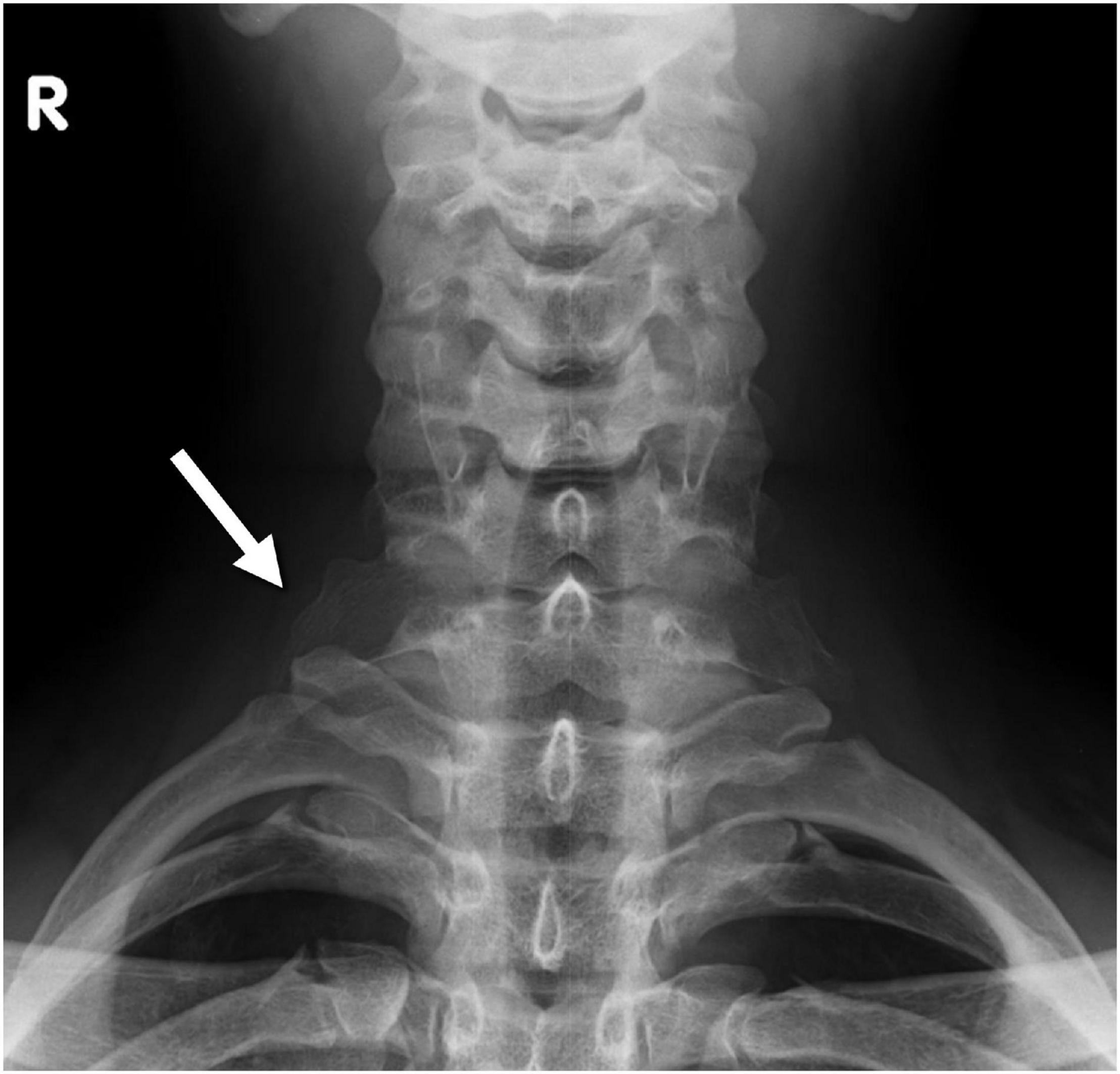
Radiographic positioning techniques for the cervical spine

Ventrodorsal projection radiograph of the thoracic vertebral

Butterfly vertebra. Coronal CT image in bone window (a) shows a
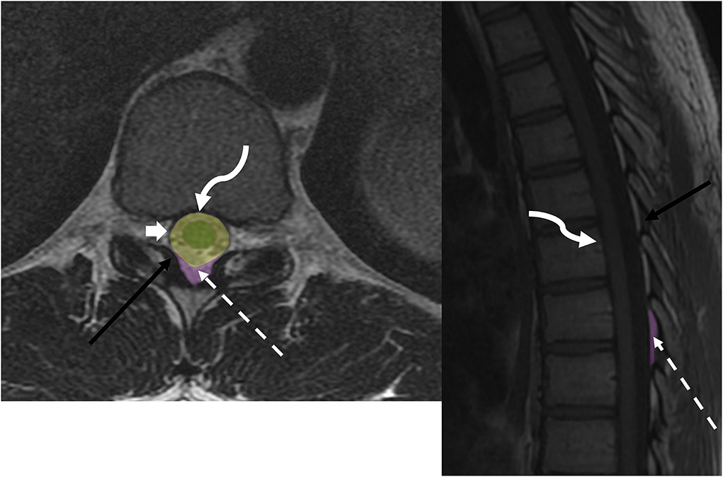
Frontiers High-Resolution Ultrasound and Magnetic Resonance Imaging of Abnormal Ligaments in Thoracic Outlet Syndrome in a Series of 16 Cases
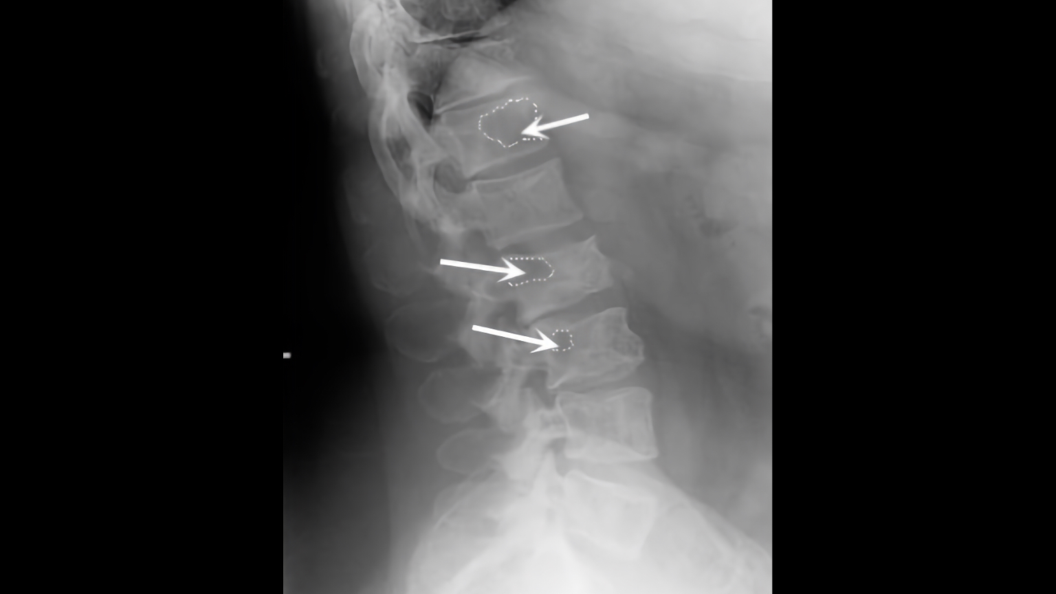
Transitional vertebra, Radiology Reference Article
Gibbus deformity, Radiology Reference Article
Lumbar X-ray Interpretation - OSCE Guide, Radiology
Frontiers Imaging of metastatic epidural spinal cord compression

Cervical and Thoracic Spine: Normal Variants and Artifacts