By A Mystery Man Writer
Structural biologist Stephen Long talks about how his team used x-ray crystallography to discover the structure of an ion channel called K2P1.

Structural Biology Memorial Sloan Kettering Cancer Center
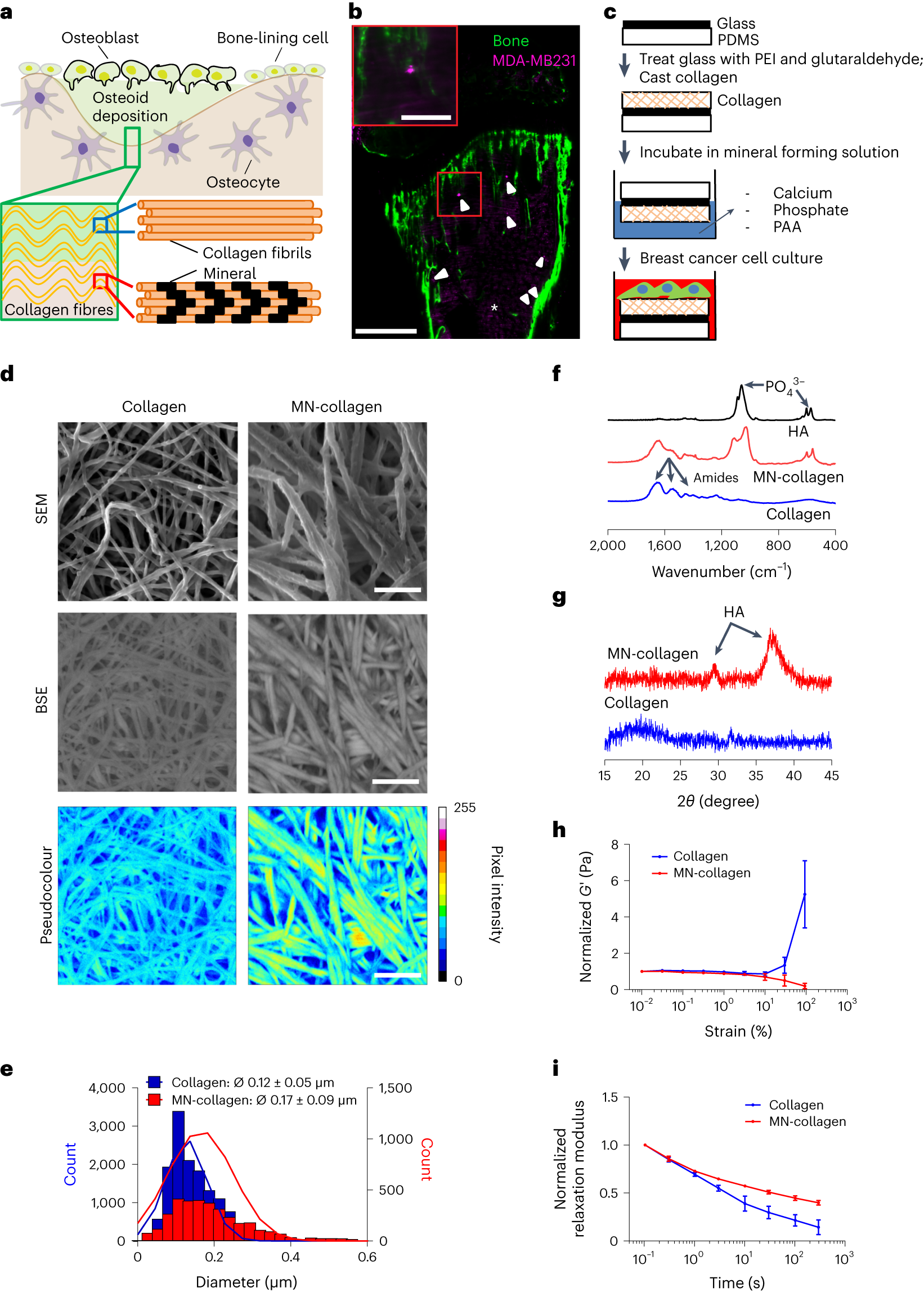
Bone-matrix mineralization dampens integrin-mediated mechanosignalling and metastatic progression in breast cancer

Memorial Sloan Kettering scoops up piece of Lipstick Building

RCSB PDB - 5T5N: Calcium-activated chloride channel bestrophin-1 (BEST1), triple mutant: I76A, F80A, F84A; in complex with an Fab antibody fragment, chloride, and calcium
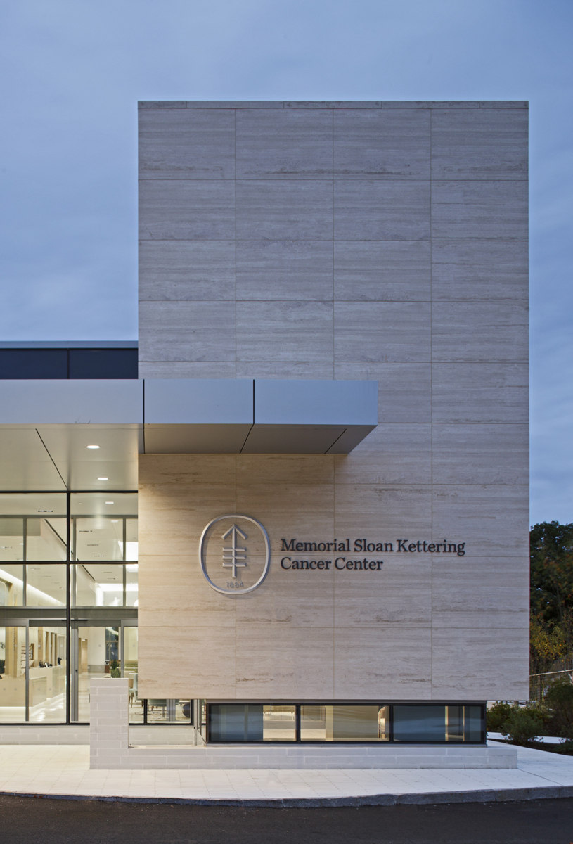
Memorial Sloan Kettering Cancer Center Westchester - Healthcare Snapshots
Memorial Sloan Kettering Cancer Center, New York, US
Quantum Leap Memorial Sloan Kettering Cancer Center

Alexandria Miller Awarded Chairman's Prize

Loss of Pip4k2c confers liver-metastatic organotropism through insulin-dependent PI3K-AKT pathway activation

Memorial Sloan Kettering Cancer Center

Memorial Sloan Kettering Hires 'Futurist' to Lead Strategic Initiatives
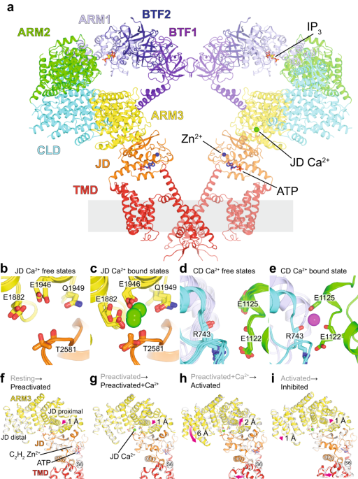
Structural titration reveals Ca2+-dependent conformational landscape of the IP3 receptor