Search
NEWS

CT image of C2–3 congenital fusion. A Vertebral body fusion and lamina

By A Mystery Man Writer

CT image of C2–3 congenital fusion. A Vertebral body fusion and lamina

Cervical osteochondroma with neurological symptoms: literature review and a case report

CT image of C2–3 congenital fusion. A Vertebral body fusion and lamina

Malformed vertebrae: a clinical and imaging review, Insights into Imaging

CT image of C2–3 congenital fusion. A Vertebral body fusion and lamina

Surgical Neurology International

CT image of C2–3 congenital fusion. A Vertebral body fusion and lamina

Cervical Myelopathy - Spine - Orthobullets

CT image of C2–3 congenital fusion. A Vertebral body fusion and lamina

The Spine: Congenital and Developmental Conditions

CT image of C2–3 congenital fusion. A Vertebral body fusion and lamina

Juedong HOU, PhD Student, Doctor of Medicine, Southern Medical University, Guangzhou, FIMMU, nanfang hospital

CT image of C2–3 congenital fusion. A Vertebral body fusion and lamina

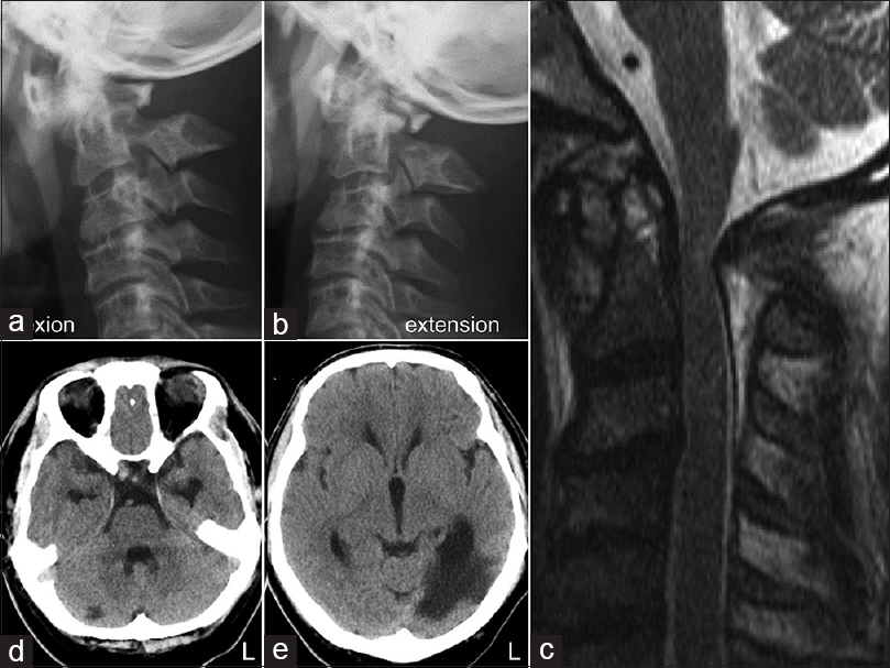
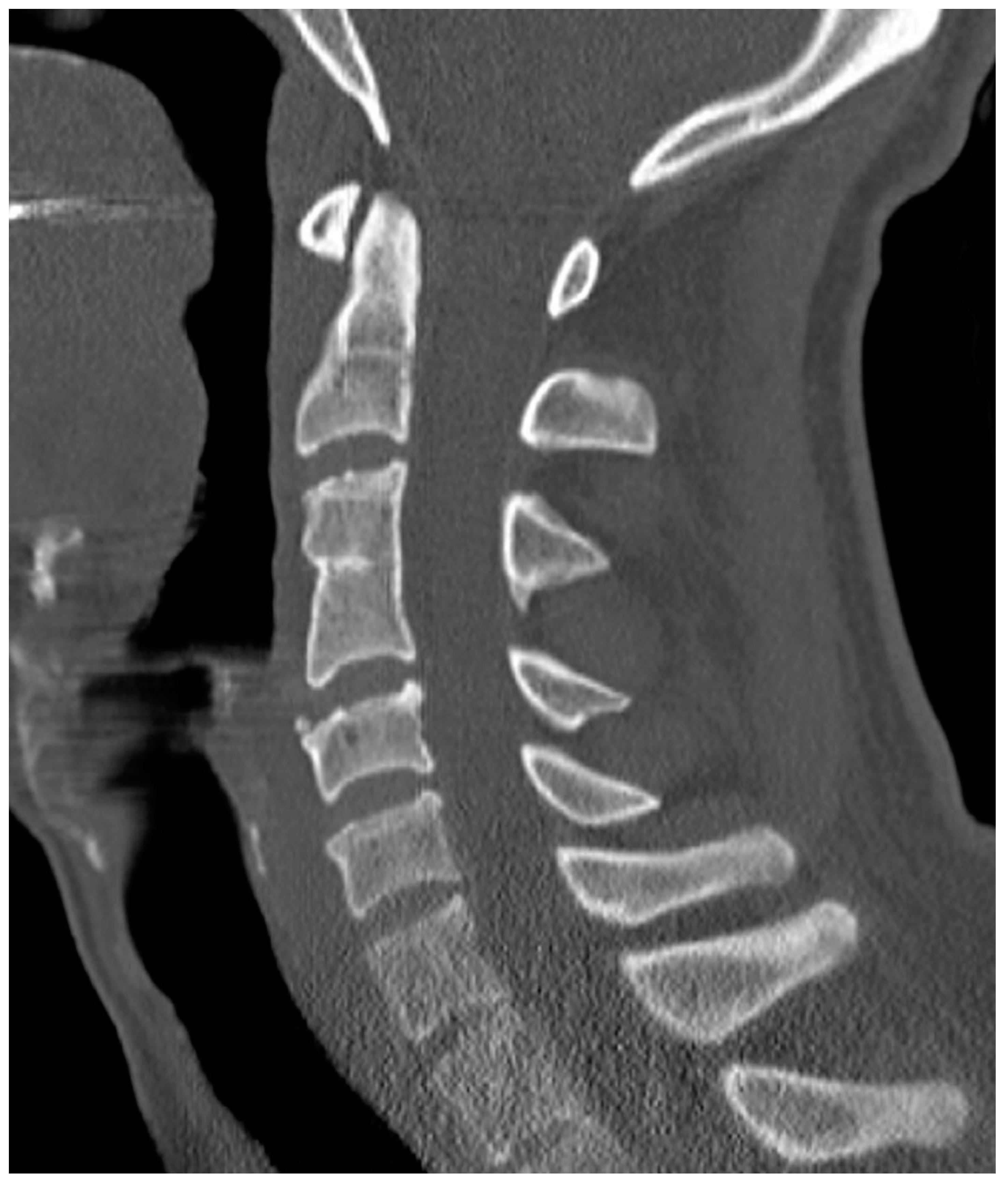
Lateral radiograph of patient with C2/C3 posterior fusion of the

CT image of C2–3 congenital fusion. A Vertebral body fusion and lamina

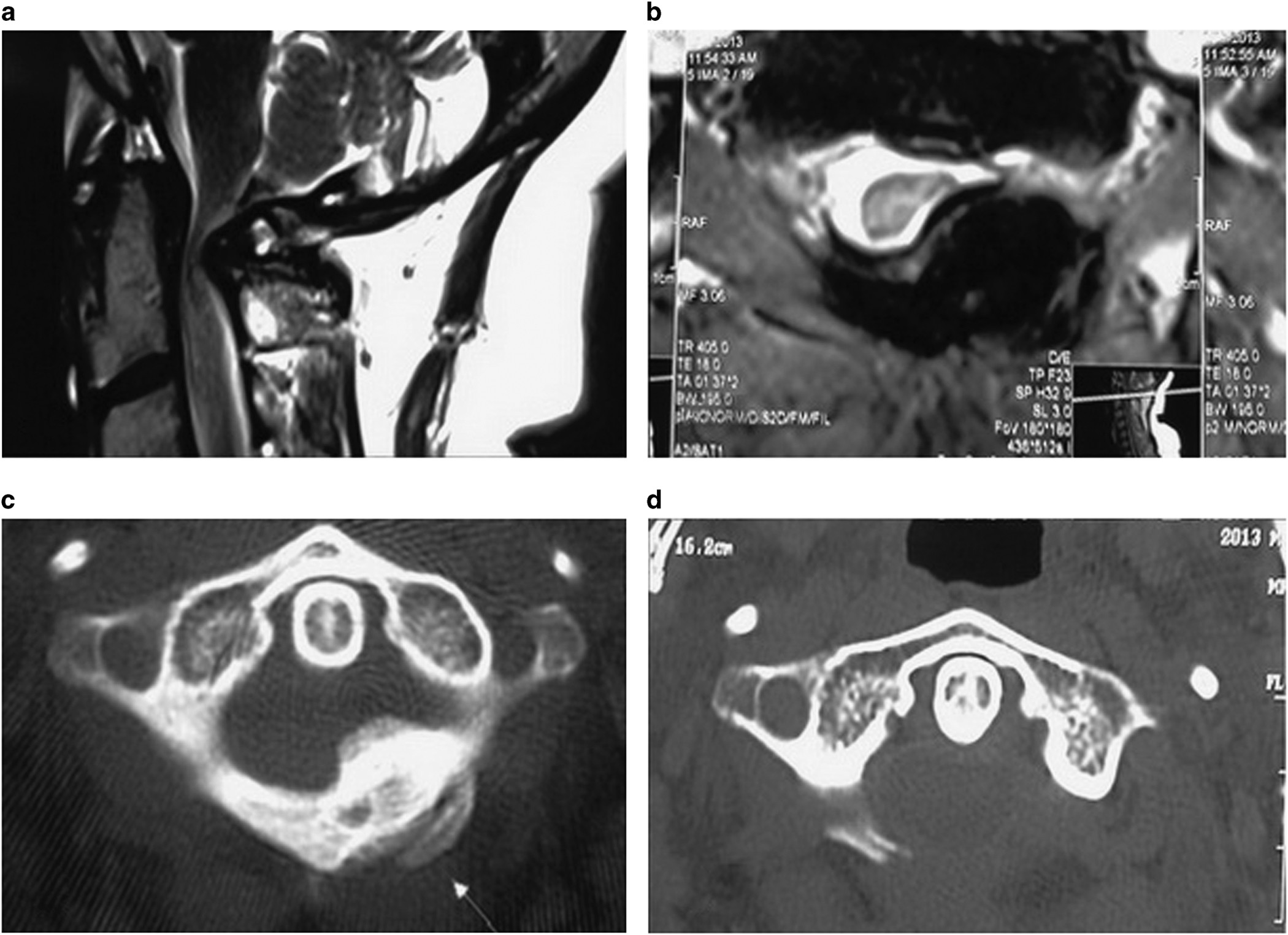
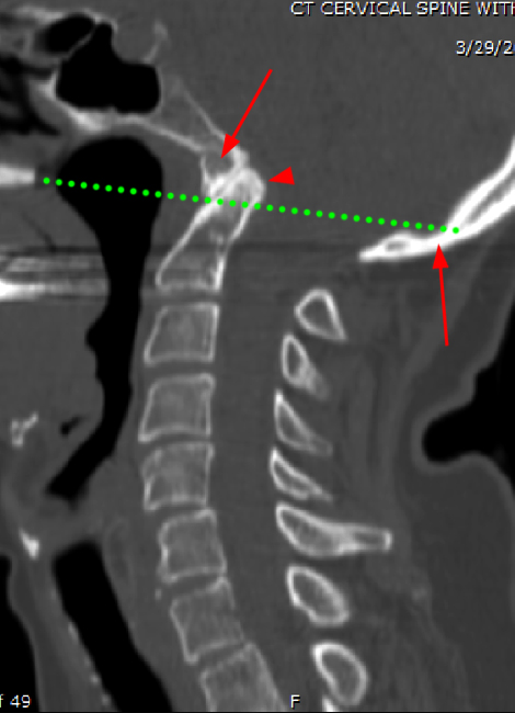
Anatomical analysis of the C2 pedicle in patients with basilar invagination

CT image of C2–3 congenital fusion. A Vertebral body fusion and lamina

EPOS™

CT image of C2–3 congenital fusion. A Vertebral body fusion and lamina

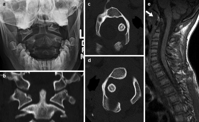
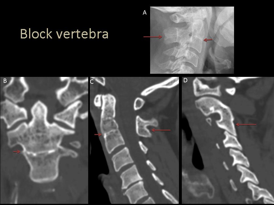
Block vertebra - C2/3, Radiology Case

CT image of C2–3 congenital fusion. A Vertebral body fusion and lamina

Klippel Feil Syndrome

CT image of C2–3 congenital fusion. A Vertebral body fusion and lamina

Diagnostics, Free Full-Text