Search
NEWS

b. Left breast USG showing oval, well-defined, mixed echogenic

By A Mystery Man Writer

b. Left breast USG showing oval, well-defined, mixed echogenic

Lactational breast changes/lobular hyperplasia mimicking masses

b. Left breast USG showing oval, well-defined, mixed echogenic

Lasya THAMBIDURAI

b. Left breast USG showing oval, well-defined, mixed echogenic

breast ultrasound Leaders in Pharmaceutical Business

b. Left breast USG showing oval, well-defined, mixed echogenic

Ultrasound Lexicon in diagnosis and management of breast

b. Left breast USG showing oval, well-defined, mixed echogenic

Complex Cystic Breast Masses: An Ultrasound Imaging Review

b. Left breast USG showing oval, well-defined, mixed echogenic

PDF) Tumoral pseudoangiomatous stromal hyperplasia: Radiological

b. Left breast USG showing oval, well-defined, mixed echogenic

Breast Ultrasound

b. Left breast USG showing oval, well-defined, mixed echogenic

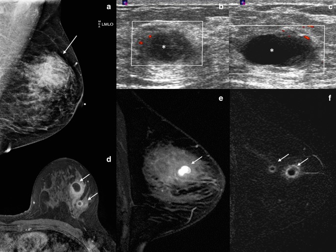
Mimickers of breast malignancy: imaging findings, pathologic

b. Left breast USG showing oval, well-defined, mixed echogenic

Hyperechoic breast images: all that glitters is not gold

b. Left breast USG showing oval, well-defined, mixed echogenic

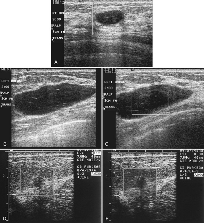
c. Left breast USG-irregular, ill-defined, multilobulated

b. Left breast USG showing oval, well-defined, mixed echogenic

b. Left breast USG showing oval, well-defined, mixed echogenic

b. Left breast USG showing oval, well-defined, mixed echogenic

Incremental value of real-time ultrasound elastography in

b. Left breast USG showing oval, well-defined, mixed echogenic

a. Case 1. Right breast USG showing well-defined, hypoechoic mass

b. Left breast USG showing oval, well-defined, mixed echogenic

b. Left breast USG showing oval, well-defined, mixed echogenic